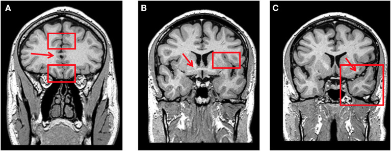
Figure 1.
(A) Coronal T1-weighted MRI at the level where the corpus callosum is first visible (red arrow). Orbitofrontal cortex is rated through the olfactory sulcus (lower red rectangle) and rostral anterior cingulate cortex through the cingulate sulcus (upper red rectangle) on this slice. (B) Coronal T1-weighted MRI at the level where the anterior commissure is first visible (red arrow). Fronto-insular cortex is rated through the circular sulcus (left-side red rectangle) on this slice and the two posterior. (C) Coronal T1-weighted MRI at the level where the connection between the frontal and temporal lobes is no more visible (left-side red arrow). Anterior temporal cortex is rated in this slice (left-side red rectangle).

