Figure 1.

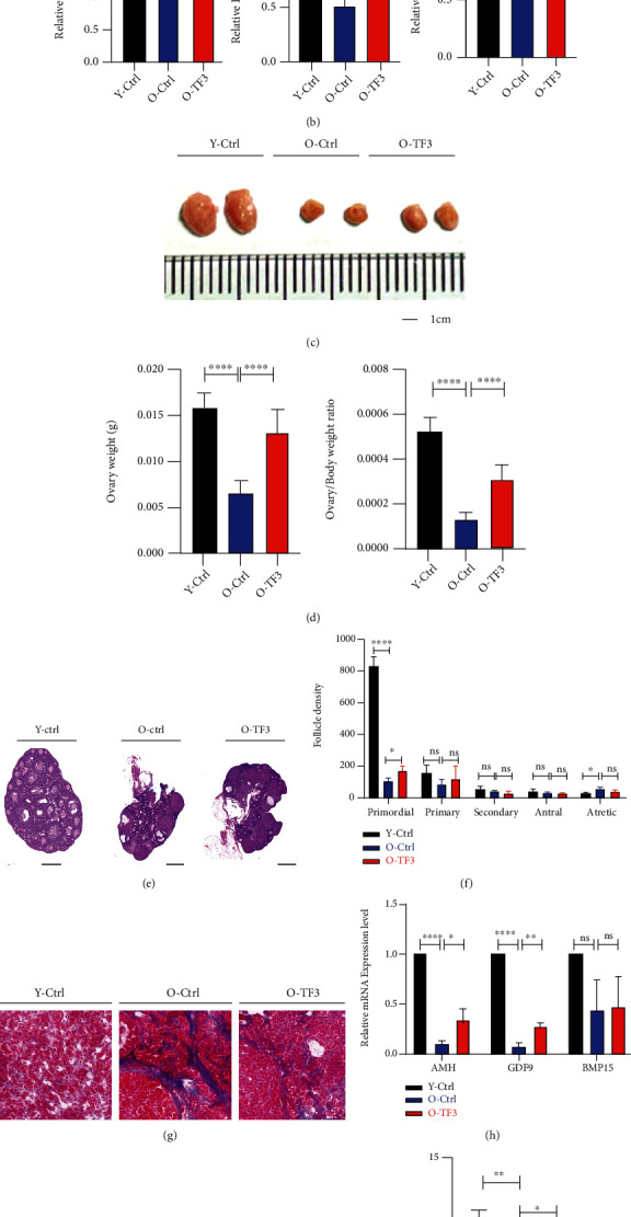
TF3 promotes ovarian function maintenance in aged mice. Vaginal cells were obtained from aged mice treated with intragastric administration (TF3 or control) for 90 days and 8-week-old young control mice without intragastric administration for 14 days. Estrous cycle was monitored and analyzed by HE staining, expressed as a line graph (a). P denotes metestrus, E denotes estrus, and M/D denotes diestrus and proestrus. Serum FSH, E2, and P4 secretion levels were measured by enzyme-linked immunosorbent assays (b). After gavage administration, the ovaries were photographed to compare the change of ovarian volume (bar = 1 cm) (c). The body and ovary weights of mice in each group after treatment were obtained, and the ovary weight and ovary/body weight ratio (d) of mice in each group were compared and analyzed. HE staining was performed on the largest cross sections to observe and analyze ovarian and follicular development (bar = 500 μm) (e). Follicle count (f) after HE staining of serial ovarian sections and Masson staining of frozen ovarian sections were performed to analyze ovarian fibrosis status (bar = 20 μm) (g). Total RNA was extracted from mouse ovaries, and the expression levels (h) of ovarian reserve markers AMH, GDF9, and BMP15 mRNA were analyzed by real-time PCR. GAPDH was the internal reference gene. At the end of gavage treatment, the mice were naturally mated with adult male mice, and the litter size (i) was recorded. Young Ctrl (Y-Ctrl): 8-week-old CD-1 mice; old Ctrl (O-Ctrl): 9-month-old CD-1 mice treated with saline for 90 days; old+TF3(O-TF3): 9-month-old CD-1 mice treated with 30 mg/kg/day TF3 for 90 days. ∗p < 0.05, ∗∗p < 0.01, ∗∗∗p < 0.001, and ∗∗∗∗p < 0.0001.
