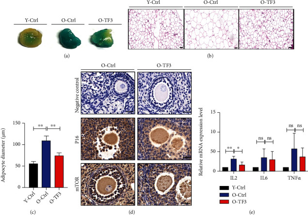
Figure 2.

Effect of TF3 on the expression of marker genes of aging in mice. After treatment, adipose tissue around the ovary was stained for the aging marker galactosidase (a) and HE to statistically analyze lipid droplet size (b, c) (bar = 50 μm). Immunohistochemical staining of ovarian tissue sections showed expression of P16 and mTOR protein, with negative control (d) on the left side (bar = 20 μm). Total RNA was extracted from the ovaries and reversely transcribed, and the relative expression levels of IL-2, IL-6, and TNFα were analyzed using real-time PCR, with GAPDH as the internal reference gene (e). ∗p < 0.05, ∗∗p < 0.01, and ∗∗∗∗p < 0.0001.
