Figure 3.

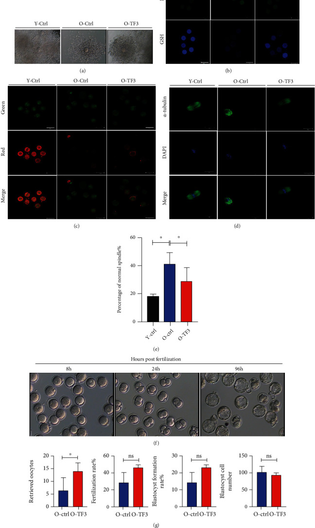
TF3 slows down the age-related decline in mouse oocyte quality. Mice in each treatment group were intraperitoneally injected with 10 IU PMSG and, after 48 h of treatment, injected with 10 IU hCG, and COCs were obtained from the ampulla of the fallopian tube 14 h later to analyze the effect of TF3 on COC morphology (a). The oxidative stress status of oocytes was analyzed by ROS and GSH staining after removing granulosa cells from COCs obtained from each treatment group (b). JC1 staining was performed to analyze oocyte mitochondrial membrane potential; red (polymers) and green (monomers) represent higher and lower oocyte mitochondrial membrane potential, respectively (c). The effect of TF3 on oocyte spindle morphology was analyzed by staining oocytes with α-tubulin antibody (green) and DAPI (blue); the right panel shows statistical analysis of abnormal spindle morphology and the ratio (d, e). COCs obtained by ovarian hyperstimulation were inseminated in vitro to analyze the effects of TF3 on oocyte retrieval, fertilization rate, blastocyst formation rate, and blastocyst cell number in aged mice (f, g). ∗p < 0.05.
