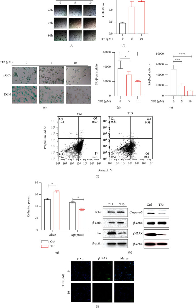
Figure 4.

TF3 inhibits apoptosis of granulosa cells cultured in vitro. Granulosa cells were routinely cultured and treated with different concentrations of TF3 (0, 5, and 10 μM) with the time to start TF3 addition set at 0 h, bar = 50 μm (a). CCK8 cell viability assay kits were used to detect the effect of TF3 on cell viability after 120 h of culture (b). Human pGCs and KGN granulosa cell lines were cultured to analyze the effect of TF3 on beta-galactosidase expression, bar = 50 μm (c). Quantitative statistical analysis of beta-galactosidase staining in pGCs (d) and KGN (e) cells was performed using ImageJ software. Flow cytometry was performed to analyze the effect of TF3 (10 μM) on pGC apoptosis (f), and the ratios of apoptosis of different types of cells were statistically analyzed (g). Total protein was extracted from pGCs treated with TF3/control, and the expression levels of apoptosis-related proteins Bcl-2, BAX, caspase-3, and γH2AX were analyzed by western blotting (h). Immunofluorescence staining was performed to analyze the expression of the apoptotic marker γH2AX, bar = 50 μm (i). ∗p < 0.05, ∗∗∗p < 0.001, and ∗∗∗∗p < 0.0001.
