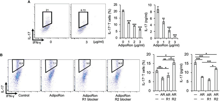
Figure 2.
AdipoRon suppresses Th17 cell differentiation via AdipoR2. (A) C1qtnf3 –/– naïve CD4+ T cells were cultured under Th17-polarizing conditions in the absence or presence of AdipoRon (0, 1, 2 and 3 μg/ml) for 5 days (n = 4 wells each). Intracellular IL-17 expression was estimated by flow cytometry after PMA/ionomycin stimulation. The number in each panel indicates percentage of IL-17+CD4+ T cells (2 left panels). The population of IL-17+CD4+ T cells (center). IL-17 concentrations in culture supernatant determined by ELISA (right). Average and SEM are shown. **p < 0.01 and ***p < 0.001. Student’s t-test. (B) C1qtnf3 –/– naïve CD4+ T cells were cultured under Th17-polarizing conditions in the absence or presence of AdipoRon (1 μg/ml, Ron) for 4 days (n = 3 wells each), and the effect of AdipoR1 (10 μg/ml, R1) or AdipoR2 blocker (10 μg/ml, R2) on the Th17 cell differentiation was examined. Intracellular IL-17 expression was estimated by flow cytometry after PMA/ionomycin stimulation. The Number in each panel indicates percentage of IL-17+CD4+ T cells (4 left panels). The population of IL-17+CD4+ T cells (center). IL-17 concentrations in culture supernatant determined by ELISA (right). Average and SEM are shown. *p < 0.05, **p < 0.01 and ***p < 0.001. Student’s t-test. All data were reproduced in another independent experiment with similar results.

