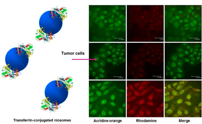
Figure 5.

Confocal microscopic analysis of intracellular localization of unmodified (L64/Chol (top), L64/Chol-R (middle)) and Tf-conjugated niosomes (L64/Chol-R-Tf (bottom)) in MCF-7 human breast cancer cells. Fluorescence of rhodamine is excited at 555 nm and detected at a wavelength of 580 nm; for acridine orange, the fluorescence is excited at 460 nm and detected at a wavelength of 650 nm. Scale bars represent 21 μm. Adapted with permission from ref (20). Copyright 2013 American Chemical Society.
