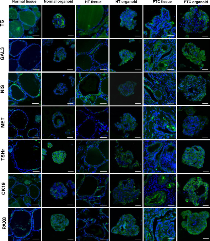
Figure 5.
Representative images of thyroids organoids subjected to immunofluorescence analysis for thyroid-specific markers, papillary thyroid carcinoma (PTC) markers. Organoids were cultured for 15 days before fixation and staining with Alexa-488 phalloidin and 4′,6-diamidino-2-phenylindole (DAPI). Scale bars, tissues = 50 µm, organoid = 20 µm. Markers indicated on the left side are shown as a green fluorescent signal. Nuclei are shown as a blue fluorescent signal.

