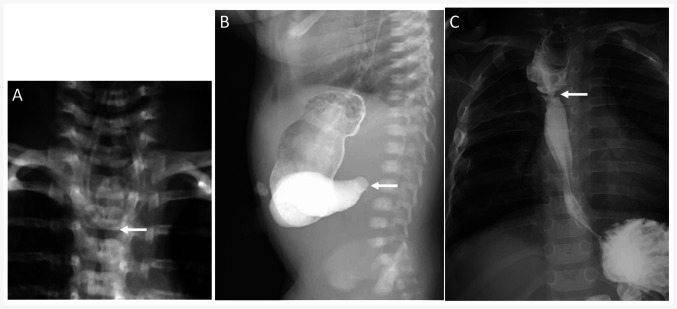
Figure 1.
X-ray analysis of patient 1. (A) A positive X-ray confirmed coiling of the nasogastric tube in the upper esophageal pouch (arrow). (B) Upper gastrography confirmed the patency of esophageal anastomosis after the first stage operation and demonstrated a large dilated stomach with duodenal obstruction (arrow). (C) Recurrent anastomotic stricture in the esophagus with <3-mm inner diameter was observed by esophageal radiography at 12 months during the follow-up period (arrow).

