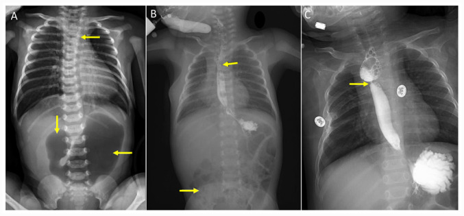
Figure 2.

X-ray analysis of patient 2. (A) A preoperative X-ray film showed the combined coiling of the nasogastric tube in the upper esophageal pouch (yellow arrows) and a large gastric bubble with no distal bowel gas (yellow arrows). (B) Upper gastrointestinal imaging confirmed patency of esophageal (yellow arrows) and duodenal anastomosis with distal bowel gas (yellow arrow). (C) Esophageal radiography confirmed esophageal anastomotic stoma stenosis with <2-mm inner diameter at 15 months during the follow-up period (yellow arrow).
