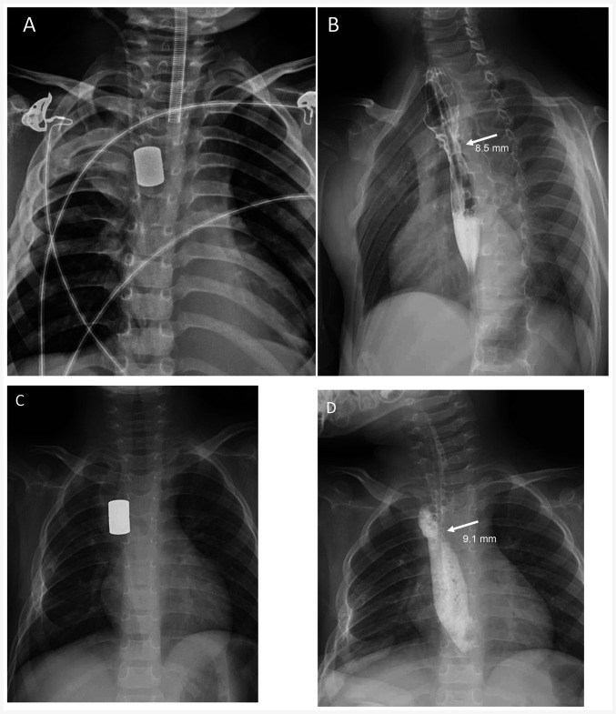
Figure 4.
Chest radiography for the two patients. (A) Radiographic approximation was performed on day 1 for patient 1. (B) Magnets were removed and radiographic examination of the patient after magnetic compression stricturoplasty showed a patent esophageal lumen with an inner diameter of 8.5 mm at the anastomotic site on day 14 (arrow). (C) Radiographic approximation was performed on day 1 for patient 2. (D) Magnets were removed on day 18 and a widened lumen was maintained (an anastomotic stoma with an inner diameter of 9.1 mm; arrow).

