
FIGURE 5.
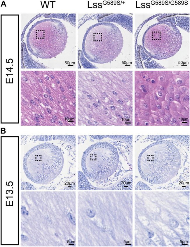
Morphology of fiber cells in embryonic lens of LssG589S/G589S mice. (A) Morphology of fiber cells stained with hematoxylin and eosin in embryonic lens of WT, LssG589S/+, and LssG589S/G589S mice at E14.5. Scale bar: 50 μm. The enlargement is the magnification of the boxed inset shown in the upper panel. Scale bar: 10 μm. (B) Morphology of fiber cells in embryonic lens of WT, LssG589S/+, and LssG589S/G589S mice at E13.5. Scale bar: 20 μm. The enlargement is the magnification of the boxed inset shown in the upper panel. Scale bar: 5 μm.