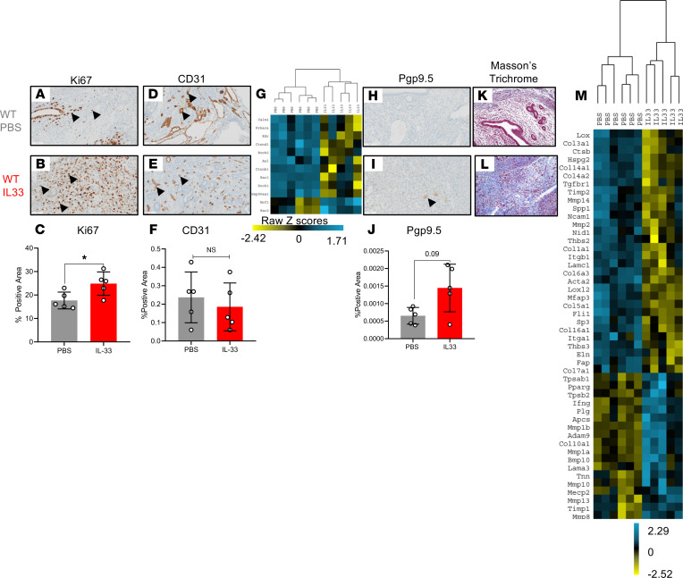
Figure 4. IL-33 alters EMS lesion proliferation, angiogenesis, innervation, and fibrosis.
(A and B) Representative images of immunohistochemical staining using Ki67, a marker for proliferation, in PBS-treated (n = 5) and IL-33–treated (n = 5) mouse lesions. (C) Quantitative analysis of the percentage of positive area of Ki67+ cells. (D and E) Representative images of immunohistochemical staining using CD31, the marker for angiogenesis, in PBS- and IL-33–treated mouse lesions, respectively. (F) Analysis of the CD31+ area percentage in lesions from PBS- and IL-33–treated mice. (G) Unsupervised hierarchical clustering using Euclidean distance and complete linkage of angiogenesis-associated genes in RNA isolated from lesions of PBS- and IL-33–treated mice. (H and I) Representative images of immunohistochemical staining using the marker for innervation, Pgp9.5, in PBS- and IL-33–treated mouse lesions. (J) Analysis of the percentage of Pgp9.5+ area. The P value (P = 0.09) for this comparison is shown. (K and L) Representative images of Masson’s trichrome staining for collagen deposition in mouse lesions from PBS- and IL-33–treated groups. (M) Unsupervised hierarchical clustering using Euclidean distance and complete linkage of fibrosis-associated genes in RNA isolated from lesions of PBS- and IL-33–treated mice. Original magnification, ×200. Scale bar: 50 μm. *P < 0.05. Mean ± SD. Nonparametric Student’s t test with Mann-Whitney.

