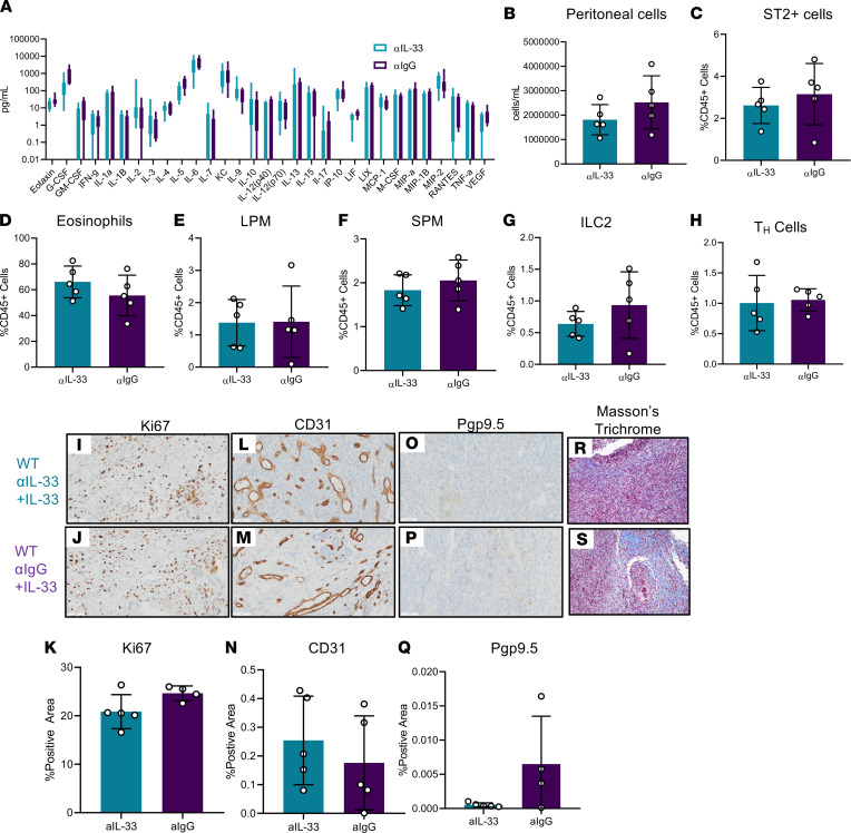
Figure 8. IL-33 Neutralization represents a promising therapeutic avenue in EMS.
WT mice were induced with endometriosis and underwent alternate day i.p. injections of IL-33 along with either a neutralizing IL-33 antibody (αIL-33) (n = 5) or IL-33 and IgG control antibody (αIgG) (n = 5). (A) Cytokine and chemokine analysis in PF samples from αIL-33– or αIgG-treated EMS mice. (B) Peritoneal lavage cells harvested from αIL-33– or αIgG-treated EMS mice counted using a Countess II FL Automated Cell Counter. (C) Frequency of ST2+ cells in the αIL-33– or αIgG-treated WT mice analyzed via flow cytometry. (D) Frequency of eosinophils (singlet, live, CD45+CD11b+F4/80+, Siglec-F+). (E) Frequency of LPM (singlet, live, CD45+CD11b+Siglec-F–F4/80hi, MHC-IIlo). (F) Frequency of SPM (singlet, live, CD45+CD11b+Siglec-F–F4/80lo, MHC-IIhi). (G) Frequency of ILC2 (singlet, live, CD45+lineage–CD25+Thy2+, ST2+). (H) Frequency of Th cells. (I–K) Representative immunohistochemical staining and quantitative analysis of lesion Ki67 staining. (L–N) Representative immunohistochemical staining and quantitative analysis of lesion CD31 staining. (O–Q) Representative immunohistochemical staining and analysis of lesion Pgp9.5 staining. (R and S) Representative histochemical staining of lesion Masson’s trichrome staining. Original magnification, ×200. Scale bar: 50 μm. Mean ± SD. Nonparametric Student’s t test with Mann-Whitney.

