
FIGURE 2.
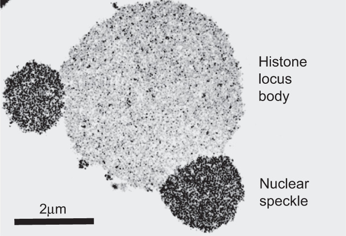
Transmission electron micrograph of two types of RNA granules within germinal vesicles of amphibian oocytes. Larger, central structure represents a histone locus body. Smaller granules fused at 5 and 9 o'clock positions upon the central histone locus body represent nuclear speckles. Scale bar = 2 µm. Photograph reproduced from Gall (2000).