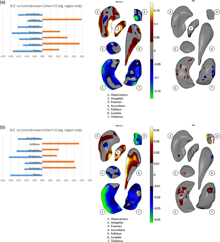
FIGURE 2.

Overall and vertex‐wise effects of diagnosis (i.e., schizophrenia vs. control) across hemispheres for (a) thickness, (b) surface dilation/contraction (log Jacobian determinant). The effects are tested in models that included diagnosis sex, age, age x sex, age2, age2 x sex, and ICV. In the left column, mean positive effect sizes and mean negative effect sizes across each subcortical structure surface (see text) are shown as bar plots. The middle column shows the vertex‐wise effects of diagnosis on interhemispheric means (see text). The right column shows the vertex‐wise effects of diagnosis on interhemispheric absolute differences (reproduced from Figure 3a,b right columns). The subcortical structures—1. hippocampus, 2. amygdala, 3. putamen, 4. accumbens, 5. pallidum, 6. caudate, and 7. thalamus—are positioned generally from a bottom viewpoint, with some slightly rotated about their own principal axis to be oblique, for better exposure: caudate—pi/7 or about 25°, accumbens—pi/10 or 18°, pallidum—pi/3 or 60°. Color scale indicates the intensity of effect sizes. Cooler colors (i.e., negative effect sizes) indicate reduced asymmetry for schizophrenia as compared to controls, and warmer colors (i.e., positive effect sizes) indicate exaggerated asymmetry. Gray color indicates nonsignificant surface vertices after multiple comparison correction
