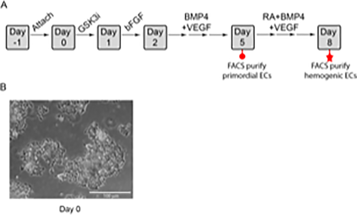
Figure 1: Protocol for the specification of primordial and hemogenic ECs.

A) Schematic diagram of the differentiation protocol. Embryonic stem cells are plated on Day −1 on matrix protein-coated plates and are allowed to attach overnight. The cells are then treated on Days 0 and 1 with GSK3i inhibitor (CHIR99021) and bFGF, respectively, to induce primitive streak and mesoderm specification, respectively. Beginning on Day 2, the cells are treated with a combination of BMP4 and VEGF-A to promote primordial EC development. Primordial ECs (red circle) are FACS purified on Day 5. Alternatively, to generate hemogenic ECs, the medium above the primordial ECs is exchanged on Day 5 to fresh hemogenic differentiation medium containing BMP4, VEGF-A, and RA. This medium is replaced daily until Day 8, when hemogenic ECs (red star) are FACS purified. B) Colonies on day 0 of differentiation, scale bar= 100 μm. Panel A has been modified from Qiu et al.37 with permission from Elsevier.
