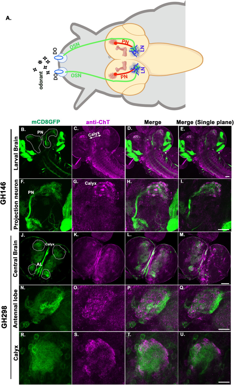
Fig 4. ChT is localised in major neurons of larval olfactory neural circuit.
(A) Schematics of the larval olfactory pathway. Odorants are detected by olfactory sensory neurons (OSNs, green), which terminate in antennal lobe (AL) and synapses with local interneurons (LNs, blue) or with dendrites of projection neurons (PNs, red). PNs innervate MB calyx (brown). (B-D) Expression of UAS-mCD8GFP in projection neurons (green) driven by GH146GAL4 and immunostained with anti-ChT (magenta) and merge. The PNs connecting AL to MB calyx are encircled by white dotted shape, scale bar 50 μm. imaged with 20x, 0.9 N.A objective (F-H) PN imaged at higher magnification, scale bar 20 μm, imaged with 40x, 1.4N.A oil objective (E and I) represents the merged image of single plane taken from the same z-section of represented image stacks. The colocalised regions can be viewed as white areas in the merged image shown. (J-M) Expression of UAS-mCD8GFP in central brain region (green) driven by GH298GAL4 and immunostained with anti-ChT (magenta) and merge, scale bar 50 μm. imaged with 20x, 0.9 N.A objective. GH298 shows expression in both AL and calyx. (N-P) AL imaged at higher magnification, scale bar 20 μm, imaged with 40x, 1.4N.A oil objective (R-T) MB calyx imaged at higher magnification, scale bar 20 μm, imaged with 40x, 1.4N.A oil objective. (M, Q, and U) represents the merged image of single plane taken from the same z-section of represented image stacks. The colocalised regions in AL and calyx can be viewed as white areas in the merged image shown. All images are z-stacked (unless mentioned) pseudocoloured representative of 3–5 3rd instar larval brains.

