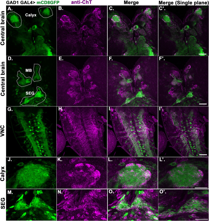
Fig 7. ChT is localised in GABAergic neurons of several neuropilar areas of Drosophila larval brain.
(A-O’) Shows 3rd instar larval brain. The pseudocolored images are mCD8GFP driven by GAD1GAL4 (green), coimmunostained with anti-ChT (magenta) and merge (colocalization appears as white). (A-F’) ChT staining marks several neuropilar areas of the central brain colocalised with areas marked by GAD1>mCD8GFP including MB, MB calyx, and subesophageal ganglia (SEG). Respective regions of central brain are encircled with white dots and labelled, scale bar 50μm, (G-I) shows VNC, scale bar 50μm, (J-L) shows high magnification image of MB calyx, scale bar 20μm. ChT staining was observed to be colocalised with GAD1>mCD8GFP marked areas, specifically at the peripheral areas of calyx. Merged image shows colocalised area as white. (M-O) shows high magnification image of SEG, scale bar 20μm. ChT staining was observed to be colocalised with GAD1>mCD8GFP marked areas, specifically at the peripheral areas of SEG. Merged image shows colocalised area as white. (C’, F’, I’, L’ and O’) represents the merged image of single plane taken from the respective z-section of represented image stacks. All images are z-stacked (unless mentioned) representative of 3–5 3rd instar larval brains.

