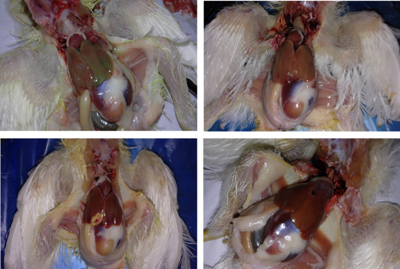
Fig 2. Gross lesions of liver in infected chickens.

a: hypertrophied greenish liver of infected chicken at 9 dpi with; b: Pallor and enlargement in liver of infected chicken at 13 dpi with; c: Necrosis lesion in liver at 13 dpi; d: Normal liver of uninfected chicken.
