
FIGURE 6.
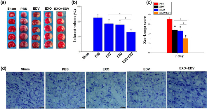
Neuroprotective role of EXO, EDV and EXO + EDV at 7 days on PMCAO rats. (a) EXO + EDV treatment reduced infarct volume. Representative brain slices with infarcts stained by 2,3,5‐triphenyltetrazolium chloride (TTC) from each group at 7 days on PMCAO rats. (b) Infarct volume on PMCAO model treated with EXO, EDV and EXO + EDV at 7 days on PMCAO rats. Data are expressed as means ± SD (n = 3). *P < 0.05, comparing infarct volume on PMCAO model treated with PBS. + P < 0.05, comparing infarct volume on PMCAO model treated with EDV with that treated with EXO + EDV. # P < 0.05. (c) Zea‐Longa neurological scores. Data are expressed as means ± SD (n = 3). *P < 0.05, comparing score on PMCAO model treated with PBS. + P < 0.05, comparing score on PMCAO model treated with EDV with that treated with EXO + EDV. # P < 0.05. (d) Representative Nissl staining of ischemic brain tissue treated with EXO, EDV and EXO + EDV at 7 days on PMCAO rats. The scale bar is 50 μm and applies to all figure parts