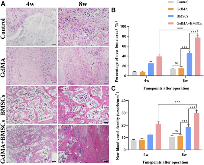
FIGURE 4.
In vivo sample staining results. (A) Histological analysis of bone defects repaired by each group at weeks 4 and 8 after surgery (bar = 200 μm). (B) The percentage of the new bone area and (C) the density of neovascularization in the repaired bone defect area in each group at weeks 4 and 8 after surgery. ***p < 0.001.

