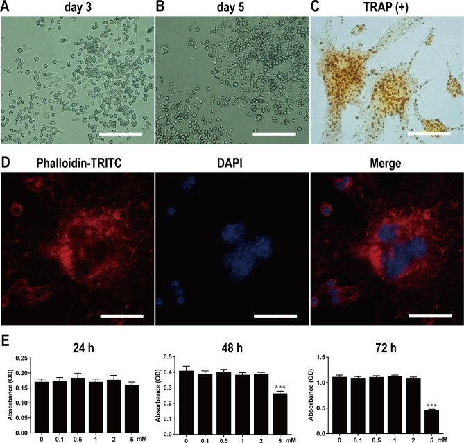
Fig 2. Isolation and culture of primary BMMs, and osteoclast differentiation, identification, and proliferation.
Light microscopy of primary BMMs on 3rd (A) and 5th d (B). Scale bar, 200 μm. Light microscopy of TRAP-positive osteoclasts with multiple nuclei (C). The F-actin ring structure stained with phalloidin-TRITC and DAPI was observed under LSCM (D). Scale bar, 40 μm. BMMs were stimulated by various concentrations of Met for 24, 48 and 72 hours, and the absorbance value was detected by CCK-8 assay at 450nm (E). n = 3 per group. ***P < 0.001, compared with control group.

