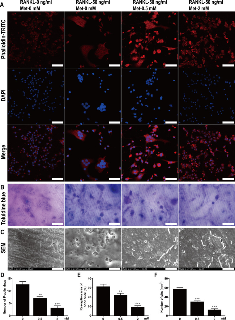
Fig 4. Met restrains F-actin rings formation and bone resorption of osteoclast in vitro.
(A) BMMs were stimulated with 30 ng/ml M-CSF, 50 ng/ml RANKL, and various concentrations of Met for 7 d and then stained with TRITC-conjugated phalloidin and DAPI to show F-actin rings and nucleus. Scale bar, 50 μm. (B) BMMs were seeded on bone slices and stimulated with 30 ng/ml M-CSF, 50 ng/ml RANKL, and various concentrations of Met for 9 d, bone resorption areas stained with toluidine blue were examined. Scale bar, 50 μm. (C) Bone resorption pits were shown by SEM. Scale bar, 20 μm. Quantitative analysis of (D) F-actin rings, (E) resorption areas and (F) resorption pits. n = 3 per group. **P < 0.01 and ***P < 0.001, compared with control group.

