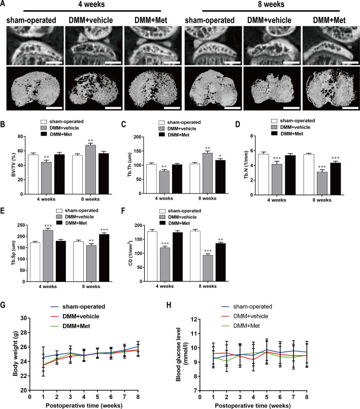
Fig 6. Met attenuates bone loss during the early stage of DMM-induced OA.
(A) The DMM-induced OA mice were treated with or without Met for 4 and 8 weeks, and then microarchitecture in tibial subchondral bone was examined by μCT. Scale bar, 1,000 μm. (B, C, D, E, and F) Quantitative μCT analyses of microarchitecture in tibial subchondral bone: (B) BV/TV (%), (C) Tb.Th, (D) Tb.N, (E)Tb.Sp and (F) CD. (G and H) Quantitative analyses of body weight and blood glucose: (G) body weight and (H) blood glucose. n = 5 per group/time point. *P < 0.05, **P < 0.01 and ***P < 0.001 compared with the sham-operated group.

