FIGURE 3.
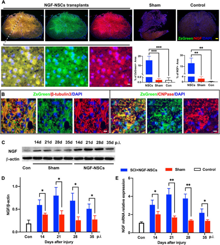
Survival and differentiation of NGF-modified NSCs after transplantation and high levels of NGF at the lesion core. (A) Four weeks after NGF-NSCs transplantation, we observed that NGF + /ZsGreen + cells aggregated in the epicenter of the injured spinal cord, and several of them migrated into the peri-lesion area. In comparison, the sham and control rats did not show a positive signal of ZsGreen, and their immunoactivity of NGF was much weaker. (B) The transplanted NGF-NSCs marked by ZsGreen expressed β-tubulins3 and CNPase, which indicated the NGF-NSCs could differentiate into neurons and oligodendrocytes. (C,D) The western blotting analysis demonstrated that NGF’s protein level was much higher in the NGF-NSCs groups at day 14, 21, 28, and 35 after injury. (E) Similarly, the mRNA level of NGF exhibited the same elevation in the three groups. Scale bar, white bar 5 μm; yellow bar 200 μm. ∗p < 0.05, ∗∗p < 0.01, and ∗∗∗p < 0.001.

