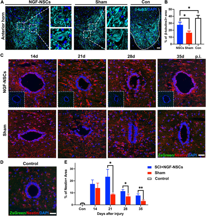
FIGURE 5.
NGF-modified NSCs transplantation protected motor neurons and enhanced endogenous neurogenesis in the peri-lesion segments. (A,B) Four weeks after NGF-NSCs transplantation, compared to the sham group, the percentage of β-tubulin3 positive area in the anterior horn was significantly higher, and the neurons maintained physiological morphology. (C) Around the central canal in the peri-lesion segments (3 mm caudal to the lesion epicenter), there were no positive signals of ZsGreen and the immunoactivity of nestin was higher in the rat receiving NGF-NSCs along the time. (D) In the control rats, the expression of nestin was weak around central canal and there were no ZsGreen + cells. (E) The semi-quantification of the percentage of nestin + area demonstrated that high expression of nestin in the peri-lesion segments of NGF-NSCs rats. Scale bar = 20 μm. *p < 0.05 and **p < 0.01.

