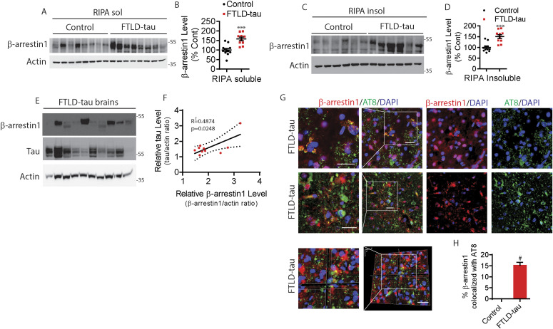
Figure 2. Elevated β-arrestin1 in FTLD-tau patient brains.
(A) RIPA-soluble extracts from the frontal cortex of healthy controls and FTLD-tau patients were immunoblotted for β-arrestin1 and actin. Representative blots are shown. (B) Quantification of RIPA-soluble β-arrestin1 levels. Healthy control (n = 12), FTLD-tau patients (n = 10). ***P < 0.0005. Unpaired t test. (C) RIPA-insoluble extracts from the frontal cortex of healthy controls and FTLD-tau patients were immunoblotted for β-arrestin1 and actin. Representative blots are shown. (D) Quantification of RIPA-insoluble β-arrestin1 levels. Healthy control (n = 12), FTLD-tau patients (n = 10). ***P < 0.0005. Unpaired t test. (E) RIPA-insoluble extracts from the frontal cortex of FTLD-tau patients were immunoblotted for β-arrestin1, tau and actin. (F) Correlation between RIPA-insoluble tau and β-arrestin1 in FTLD-tau patients (n = 10 FTLD-tau; R2 = 0.4874, P = 0.0248, linear regression). (G) Representative images and Z-stack images of FTLD-tau brains showing that β-arrestin1 and AT8+ (pS202/pT205) tau pathology are colocalized (Scale bar = 20 μm). White boxes are magnified left. (H) Quantification of colocalization between β-arrestin1 and AT8. #P < 0.0001. Unpaired t test.

