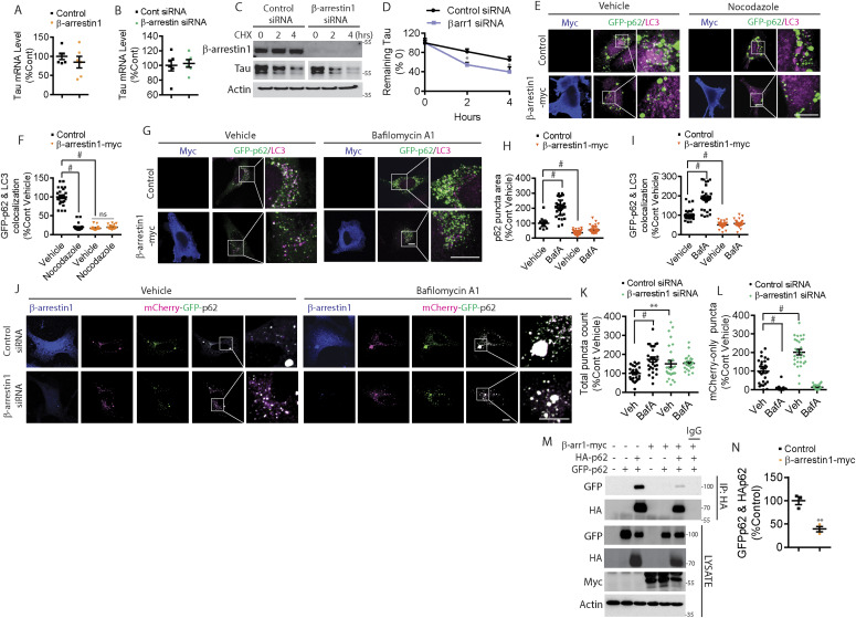
Figure 6. β-arrestin1 inhibits autophagy-mediated tau clearance.
(A) Quantification of β-arrestin1 mRNA levels by qRT-PCR in HeLa-V5-tau cells transfected with control vector or β-arrestin1. n = 6 independent experiments. (B) Quantification of β-arrestin1 mRNA levels by qRT-PCR in HeLa-V5-tau cells transfected with either control siRNA or β-arrestin1 siRNA. n = 6 independent experiments. (C) HeLa-V5-tau cells were transfected with control siRNA or β-arrestin1 siRNA and treated with cycloheximide (100 μg/ml) for 2 and 4 h. Cells were subjected to immunoblotting for β-arrestin1, tau, and actin. Representative blots are shown. (D) Quantification of tau remaining after cycloheximide treatment. n = 3 independent experiments. *P < 0.05. Two-way repeated-measures ANOVA. (E) Confocal images of HeLa-V5-tau cells transfected with GFP-p62 with either control vector or β-arrestin 1-myc and treated with vehicle or 20 μM of nocodazole for 30 min. Cells were fixed and immunostained for myc and LC3 (Scale bar = 10 μm). Representative images are shown. (F) Quantification of GFP-p62 and LC3 colocalization. n = 4 independent experiments. #P < 0.0001. One-way ANOVA with Dunnett’s post hoc test. (G) Confocal images of HeLa-V5-tau cells transfected with GFP-p62 and either vector control or β-arrestin1-myc and treated with vehicle or 100 nM of Bafilomycin A1 for 4 h. Cells were fixed and immunostained for myc and LC3 (Scale bar = 10 μm). Representative images are shown. (H) Quantification of GFP-p62 puncta area. n = 4 independent experiments. #P < 0.0001. One-way ANOVA with Dunnett’s post hoc test. (I) Quantification of GFP-p62 and LC3 colocalization. n = 4 independent experiments. #P < 0.0001. One-way ANOVA with Dunnett’s post hoc test. (J) Confocal images of HeLa-V5-tau cells transfected with mCherry-GFP-p62 with either control siRNA or β-arrestin1 siRNA and treated with vehicle or 100 nM of Bafilomycin A1 for 4 h. Cells were fixed and immunostained for β-arrestin1 (scale bar = 10 μm). mCherry is pseudocolored to magenta. Representative images are shown. (K) Quantification of total p62 puncta (mCherry+GFP) normalized to control vehicle treatment. n = 4 independent experiments. #P < 0.0001, **P < 0.005. One-way ANOVA with Dunnett’s test. (L) Quantification of mCherry-only (magenta) puncta normalized to control vehicle treatment. n = 4 independent experiments. #P < 0.0001. One-way ANOVA with Dunnett’s test. (M) HeLa-V5-tau cells were transiently transfected with control vector or β-arrestin1-myc together with either GFP-p62 and/or HA-p62 and subjected to co-immunoprecipitation for HA and immunoblotting for GFP, HA, myc, and actin. Representative blots are shown. (N) Quantification of GFP-p62 and HA-p62 interaction n = 3 independent experiments. **P < 0.005. Unpaired t test.

