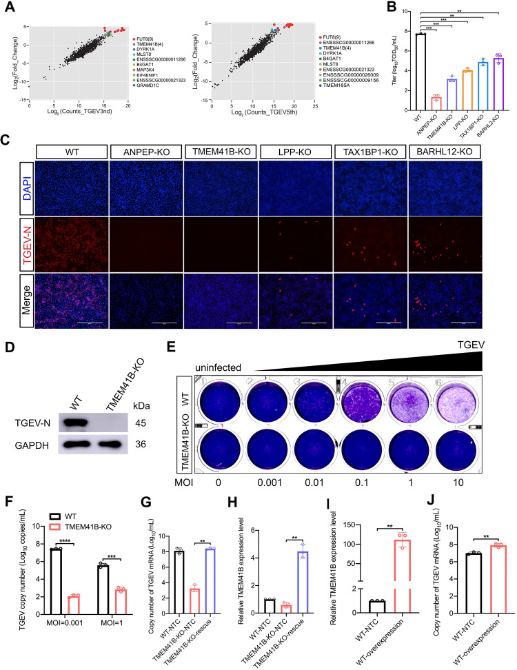
Fig 2. TMEM41B is a host factor required for TGEV replication.
(A) Verification of candidate genes enriched in the genome-wide CRISPR screen using a second, focused-CRISPR library screen. Scatter plots compare sgRNA targeted sequence frequencies and the extent of enrichment in the transformed PK-15-Cas9 cells (mock-treated versus TGEV infected) for the third (TGEV3rd) and fifth (TGEV5th) rounds of TGEV screening. Counts_TGEV3rd and Counts_TGEV5th represent the average values of the read counts from paired-end sequencing. Log2(fold change) is the median log 2 ratio between normalized sgRNA count of TGEV challenged and mock-treated cell populations. (B) Quantification of virus infectivity (TCID50) in culture supernatant collected 24 hpi from TGEV-infected (MOI = 0.1) WT and KO (ANPEP, TMEM41B, LPP, TAX1BP1, BARHL12) cell lines. (C) Immunofluorescence assays for detection of the TGEV N protein in WT cells and five selected genes (ANPEP, TMEM41B, LPP, TAX1BP1, BARHL12) KO cells following infection with TGEV (MOI = 0.1) at 24 hpi. Scale bar, 200 μm. (D) Western blot assay to detect the TGEV N protein expressed in TMEM41B KO and WT cells following infection with TGEV (MOI = 1) at 24 hpi. GAPDH used as an internal control gene. (E) Viral quantification by plaque assays. TMEM41B KO and WT cells were seeded into 24-well culture plates and infected with TGEV at different MOIs (0, 0.001, 0.01, 0.1, 1 and 10). Plates stained with 1% crystal violet to view plaques. (F) TMEM41B KO and WT cells were infected with TGEV at different MOIs (0.001 and 1), TGEV N copy number was assessed by absolute quantitative real-time PCR. (G and H) Rescue assays for WT, TMEM41B-KO and TMEM41B-KO-rescue cells infected with TGEV (MOI = 1). RT-qPCR assay for determination of (G) relative mRNA level of TMEM41B and (H) absolute mRNA level of TGEV N gene. TMEM41B-KO-rescue: reconstituted TMEM41B in TMEM41B KO cells. (I and J) Overexpression of TMEM41B in PK-15 control cells following infection with TGEV (MOI = 1). RT-qPCR assay for the determination of (I) relative mRNA level of TMEM41B and (J) absolute mRNA level of the TGEV N gene. PK-15-NTC: Transfection of pcDNA3.1 empty vector in WT cells; PK-15-overexpression: Transfection of pcDNA3.1-TMEM41B vector in WT cells. WT, wild-type; KO, knockout; MOI, multiplicity of infection; kDa, kilodaltons; DAPI, 4’,6-diamidino-2-phenylindole. **P < 0.01; ***P < 0.001; ****P <0.0001. P values were determined by two-tailed Student’s t-tests. Data are represented as a percentage mean titer of triplicate samples relative to WT control cells ± S.D.

