Fig. 8.

Phase contrast photomicrographs and graphical representation of MCF‐7 cell line
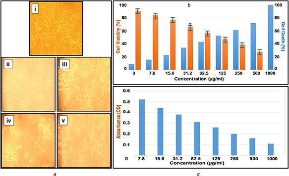
(a) Morphological changes in MCF‐7 cells treated with PtNPs. The changes in the cell morphology due to toxicity of PtNPs were determined by phase contrast microscopical analysis: (i) normal breast cancer cell line, (ii) Toxicity‐1000 µg/ml, (iii) Toxicity‐125 µg/ml, (iv) Toxicity‐62.5 µg/ml and (v) Toxicity‐31.2 µg/ml, (b) Assessment of PtNPs induced cell death in MCF‐7 breast cancer. The cells were treated with a range of concentration of PtNPs and incubated for 24 h, the per cent of cell viability and cell death was determined by MTT assay, (c) Absorbance analysis for MCF‐7 cell line with various concentrations of biological synthesised PtNPs
