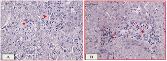
Fig. 9.

Histology observations in the healthy hepatocellular tissue model,
(a) the fibrous connective tissue surrounded the hepatocytes in portal area which is related to the negative control group with neither RF exposures nor GS@IONPs presence, (b) positive group with RF exposures without nanoparticles presence. Not significant changes can be seen in the H&E staining tissue model with ×100 magnification of light microscope. Fibrosis (►), lymphatic infiltration (7), hepatocytes (dark dots), the scale bar equals 50 µm
