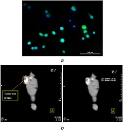
Fig. 6.

(a) Fluorescent microscopy images (20X) of dendrimer‐G2 ‐ FITC complex on MCF‐7 cells. Nuclei were stained with DAPI (blue), (b) CT images of the mice injected with dendrimer‐G2 ‐Iohexol complex 30 min after injection. The mice injected intravenously into their tail vein. Tumour size was found to be 1.07 cm
