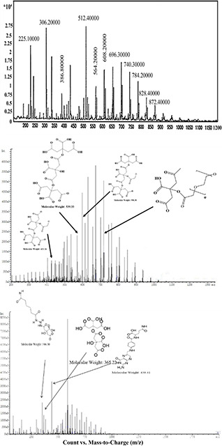
Fig. 4.

LC‐MS spectroscopy of
(a) Dendrimer‐G2, (b) Dendrimer‐G3, (c) Dendrimer‐G3‐folic acid. The peak around 306 m/z indicates the fragmentation of a citric acid attached to the second citric acid and a repetitive unit of PEG. The peak located at around 512 represents five repetitive units of PEG conjugated to the citric acid molecules and the peak around 696 m/z showed seven repetitive unite of PEG along with two citric acid molecules
