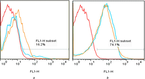
Fig. 9.

Flow cytometry analysis of endosomal escape using dequenching of carboxyfluorescein alone (negative control, red lines) in HeLa cells or following co‐incubation with
(a) 0.5 mg/ml or (b) 1 mg/ml MCM‐41‐Im (blue lines) and MCM‐41‐NH2 (orange lines)
