
Fig. 3.
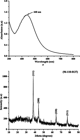
SPR of silver NPs was visualised at 440 nm and X‐ray diffraction pattern of the silver NPs matched with the standard JCPDS Ag pattern
a UV‐vis absorption spectra of the synthesised silver NPs
b XRD pattern spectra of the synthesised silver NPs