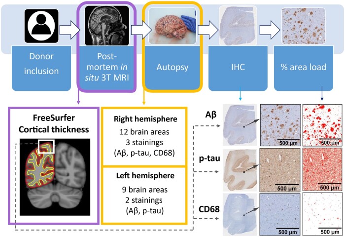
Figure 1.
Workflow of the post-mortem MRI-pathology pipeline. Once the donors were included in the study, they received a post-mortem in situ 3T MRI, and cortical thickness was calculated with FreeSurfer25 from the 3D T1w image (purple box). After the MRI scan, autopsy was performed, and brain tissue was processed for immunohistochemistry against Aβ, p-tau and CD68 (yellow boxes), which were quantified using ImageJ. The correlation between cortical thickness and %area of immunoreactivity was investigated via linear mixed models (dashed grey arrow). Aβ, amyloid-beta; IHC, immunohistochemistry; p-tau, phosphorylated-tau.

