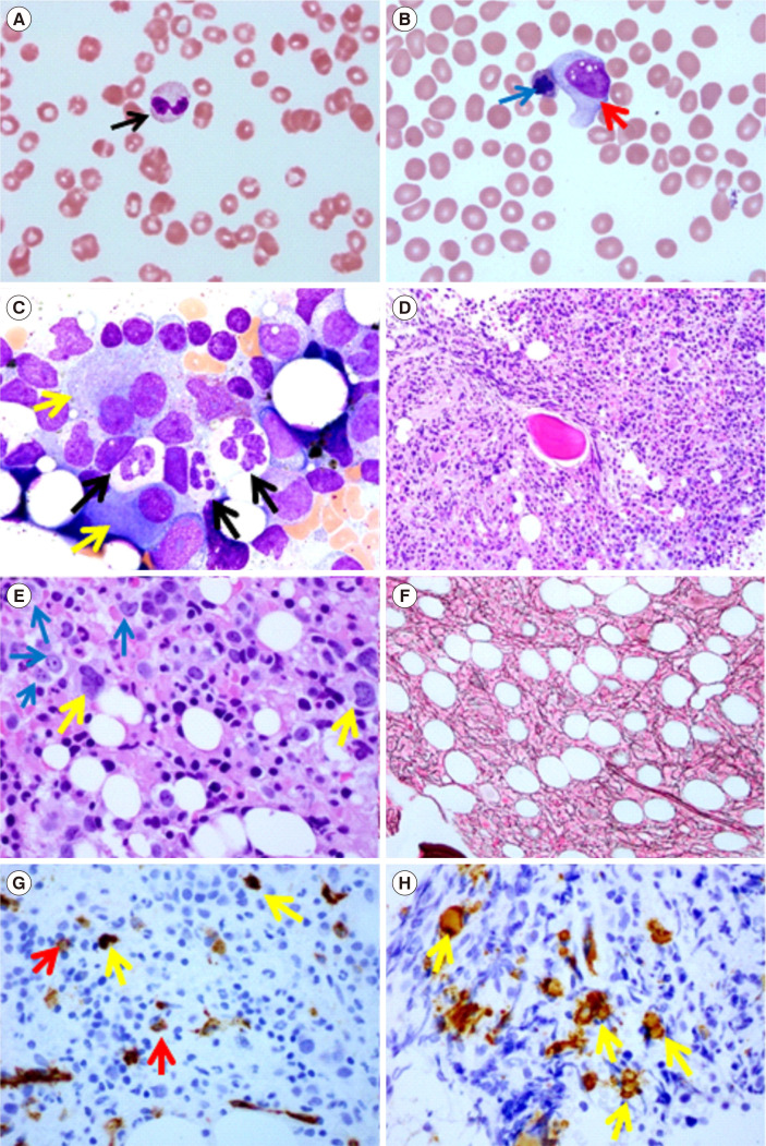
Fig. 1.
Fibrotic MDS. (A, B) Peripheral blood smears revealing dysplastic neutrophils with pseudo-Pelger–Huet changes (black arrow–bilobed neutrophil, usually indicates an underlying BM disorder) and circulating dysplastic erythroid precursors with multilobulated nuclei (blue arrow) and a circulating blast (red arrow) - leukoerythroblastosis (Wright stain, ×1,000). (C) BM touch imprint smear showing hypogranulated neutrophils (black arrows) and a bilobed megakaryocyte (yellow arrow) (Wright–Giemsa stain, ×1,000). (D, E) BM core biopsy showing hypercellularity (>90%) associated with increased left-shifted erythroid precursors (blue arrow), dysplastic megakaryocytes (yellow arrow), decreased myeloid precursor numbers, and patchy crush artifact (hematoxylin and eosin stain, ×200 [D], and ×600 [E]). (F) Reticulin stain (×200) showing moderate reticulin fibrosis (MF-2/3). (G) CD34-stained (brown colored) occasional myeloblasts (red arrows) and occasional small megakaryocytes/megakaryoblasts (yellow arrows) and (H) CD61-stained megakaryocytes, including small forms (immunoperoxidase stain, ×200).
Abbreviation: MDS, myelodysplastic syndrome; BM, bone marrow.

