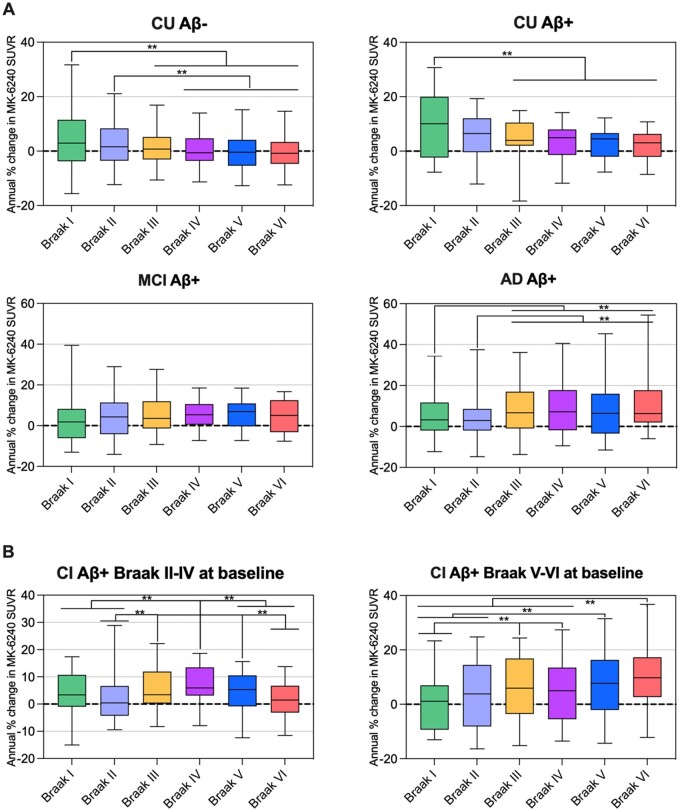
Figure 4.
Longitudinal patterns of 18F-MK-6240 SUVR accumulation in individuals across the ageing and Alzheimer’s disease spectrum. Box plots showing the quartiles, 5th and 95th percentiles (whiskers) of the annual percentage of change in 18F-MK-6240 SUVRs in different Braak-related areas in individuals segregated based on their baseline cognition, amyloid-β status and Braak stage [CU Aβ− (n = 65), CU Aβ+ (n = 22) and MCI Aβ+ (n = 21), AD Aβ+ (n = 17), CI Aβ+ and Braak II–IV (n = 20) and CI Aβ+ and Braak V–VI (n = 18)]. Statistical differences between groups were derived from the estimated differences between the means from the repeated measures analysis and the significant differences are indicated with **P < 0.05. The comparisons that survived a strict Bonferroni correction considering all the comparisons shown in the figure were: CU Aβ− (Braak I–IV, I–V and I–VI), CU Aβ+ (Braak I–IV), AD Aβ+ (Braak I–VI and II–VI), CI Aβ+ and Braak II–IV at baseline (Braak II–IV and IV–VI), CI Aβ+ and Braak V–VI at baseline (Braak I–III, I–V, I–VI and II–VI). (A) Cognitively unimpaired participants showed higher 18F-MK-6240 SUVR accumulation in PET Braak-like stage I than in the latest PET Braak-like stages, MCI Aβ+ individuals did not present differences in the rates of progression across Braak regions and AD Aβ+ patients showed lower tau increases in Braak regions I–II than in the later Braak regions. (B) We also assessed cognitively impaired individuals segregated based on their baseline Braak stage. CI Aβ+ Braak II–IV individuals at baseline showed higher tau accumulation in Braak regions III–V than in regions II and VI, whereas CI Aβ+ Braak V–VI individuals at baseline showed higher tau accumulation in Braak VI than in Braak regions I–IV. CI = cognitively impaired with MCI or Alzheimer’s disease dementia.

