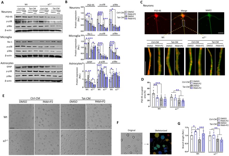
Figure 4.
Response of primary neurons, microglia, astrocytes and neuron-astrocyte co-cultures to Tat and PAM. (A) Primary neurons, microglia, astrocytes were isolated from 1-day-old pups of wild-type (Wt) and α7−/− mice, treated with the conditioned medium from pcDNA3-transfected cells (Ctrl-CM) or pcDNA3-Tat-transfected cells (Tat-CM) and PNU-125096 (PAM, 1 µM) and an α7 agonist PNU-282987 (P2, 0.5 µM) for 24 h and harvested to determine expression of PSD-95, Iba-1 or GFAP, or p-p38, p38 and β-actin by western blotting. (B) Protein expression was quantitated as in Fig. 1E. (C) Primary neurons were also double immunostained for PSD-95 and MAP2. (D) The positive staining area of PSD-95 puncta was quantitated by ImageJ. Primary microglia were visualized for their morphologies by microscopy before harvesting for cell lysates (E) and then skeletonized (F). The line shaped branches, indicative of the ramified stage, and their total length are quantified in G. The cells with shorter, non-completely formed or no line shaped branches were recognized as more amoeba-like phenotypes, which presented more often in only Tat treated wild-type group and α7- with Tat treatment groups (A). Multiple independent repeats were used for statistical analysis (n = 3/group for western blotting; n = 8/group for immunofluorescent staining, n = 6/group for microglia morphology). P < 0.05 was considered significant and is denoted with an asterisk; **P < 0.01 and ***P < 0.001 were considered highly significant. Scale bars = 10 µm (C) and 50 µm (E).

