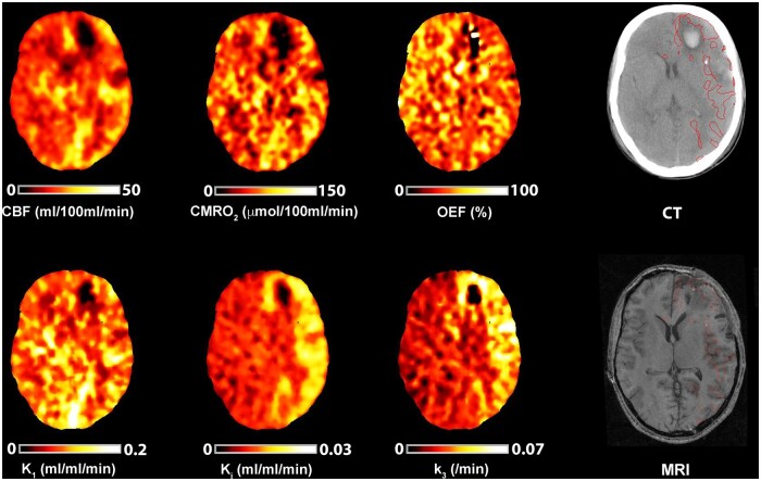
Figure 6.
Association between early metabolic derangements and tissue fate. Imaging obtained in a 17-year-old male with moderate TBI following a road traffic accident with initial GCS of 12, but who required sedation and ventilation for control of raised ICP. Co-registered CBF, CMRO2, OEF, 18F-FDG kinetic parameters and CT imaging obtained on Day 8 following TBI. Plasma glucose during imaging was 5.4 mmol/l. The CT demonstrates left frontal and temporal haemorrhagic contusions and is shown with the T1-weighted MRI obtained at 9 months post-injury; the MRI has been non-rigidly registered to the CT. Both structural images are displayed with the region of k3 hotspot outlined in red, but both lesion core and penumbra have been excluded from the hotspot to ensure that only increases in k3 outside of the lesion identified on CT imaging are shown. The volume of brain within the k3 hotspot in this subject was 149 ml and, in comparison with brain that appeared structurally normal, had CBF 19.6 versus 19.6 ml/100 ml/min, CMRO2 55.2 versus 60.2 µmol/100 ml/min, OEF 37.8 versus 42.4%, K1 0.097 versus 0.087 ml/ml/min, Ki 0.018 versus 0.012 ml/ml/min and k3 0.058 versus 0.024/min, respectively. The T1-weighted MRI demonstrates established lesions within the left frontal and temporal brain regions in close proximity with k3 increases outside of lesion core and penumbra identified on the CT image.

