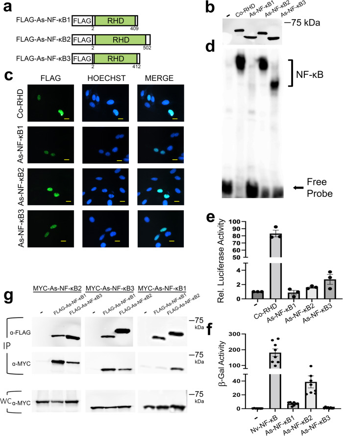
Fig. 6. Characterization of cellular and molecular properties of three choanoflagellate NF-κBs.
a FLAG-tagged NF-κB proteins used in these experiments. From top to bottom the drawings depict the three NF-κB-like proteins from the transcriptome of A. spectabilis5. RHDs are in green (Rel Homology Domain). b Anti-FLAG Western blot of lysates from HEK 293T cells transfected with the indicated expression vectors or the vector control (-). Raw image is in Supplementary Fig. 13. c Indirect immunofluorescence of DF-1 chicken fibroblasts transfected with the indicated expression vectors. Cells were then stained with anti-FLAG antiserum (left panels) and Hoechst (middle panels), and then merged (right panels). Yellow scale bar is 10 µm. d A κB-site electromobility shift assay using a palindromic κB-site probe (GGGAATTCCC) and each of the indicated lysates from b. The NF-κB complexes and free probe are indicated by arrows. Raw image is in Supplementary Fig. 13. e A κB-site luciferase reporter gene assay was performed with expression vectors for the indicated proteins or the empty vector control (-) in HEK 293 cells. Luciferase activity is relative (Rel.) to that seen with the empty vector control (1.0). Values are averages of n = 3 biological replicates per sample each performed in triplicate, and are reported with standard error. Raw data are in Supplementary Data 5. f A GAL4-site LacZ reporter gene assay was performed with the indicated GAL4-fusion proteins or the GAL4 alone vector control (-) in yeast Y190 cells. Values are average β-gal units of n = 8 biological replicates. Raw data are in Supplementary Data 5. g Co-immunoprecipitation (IP) assays of MYC-tagged As-NF-κB1, As-NF-κB2, As-NF-κB3. In each IP assay, MYC-As-NF-κBs were co-transfected in HEK 293T cells with the pcDNA-FLAG vector control (-), or FLAG-As-NF-κB1, 2 or 3 as indicated. An IP using anti-FLAG beads was performed, and pulled down proteins were then analyzed by Western blotting with anti-FLAG (top) or anti-MYC (middle) antisera. An anti-MYC Western blot of 5% of the whole-cell (WC) lysates used in the pulldowns was also performed (bottom). Raw image is in Supplementary Fig. 13.

