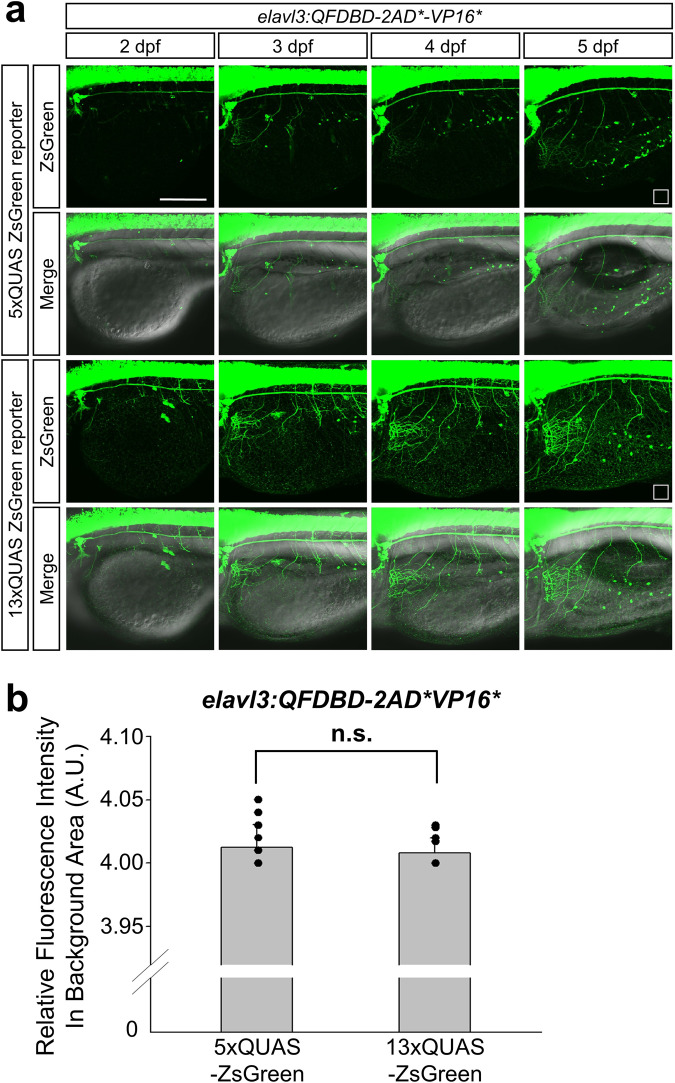
Fig. 6. The intensity of ZsGreen was gradually mounting on the sensory neurons in EYL across embryonic growth.
a Representative image of embryos at the indicated developmental stage. They obtained from genetic crosses between F2 Tg(elavl3:QFDBD-2xAD*-VP16*) and F3 effector lines having 5x or 13xQUAS-regulated ZsGreen reporter gene. b The points labeled with white open square box in (a) stand for the region used to level off background fluorescence intensity of 5x and 13xQUAS-ZsGreen groups. Comprehensive level of background fluorescence was measured with 12 single images derived from same number of embryos at 5 dpf under the identical confocal setting. No discernable differences were observed in two discrete clutches. Twelve embryos in each clutch were analyzed in the assay. Abbreviations: A.U.; arbitrary unit, n.s.; not significant, Teb; tebufenozide. Scale bar: 200 μm.

