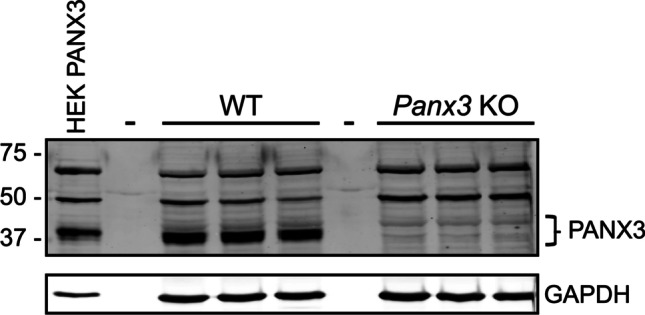
Fig. 2.

Both 50 kDa and 70 kDa immunoreactive species are seen in both WT and Panx3 KO mouse tissue when immunoblotting with the anti-PANX3 CT-379 polyclonal antibody. Western blots using WT and Panx3 KO postnatal day 5 hindlimb protein lysates were probed with a 1:1000 dilution of anti-PANX3 CT-379 antibody. The 43 kDa PANX3 species is only seen in WT tissue but multiple immunoreactive species at 50 kDa and 70 kDa are seen in hindlimbs of both genotypes. Dashes represent empty lanes. WT, wildtype. KO, knockout. HEK PANX3, human embryonic kidney cells (HEK-293 T), ectopically expressing a mouse PANX3 plasmid. N = 3 per genotype. GAPDH, glyceraldehyde 3 phosphate dehydrogenase was used as a protein loading control. Protein sizes in kDa
