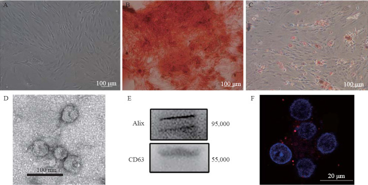
Figure 1.

Identification of MSCs and MSC-Exo. A: the morphology of the MSCs; B: differentiation of MSCs into osteoblasts; C: differentiation of MSCs into adipocytes; D: electron microscopic image of exosomes; E: Western blotting luminograms showing exosomal expressions of CD63 and Alix; F: PKH26-tagged (red fluorescence) exosomes in the neuronal cytoplasm. MSCs: mesenchymal stem cells; MSC-Exo: MSC-derived exosomes.
