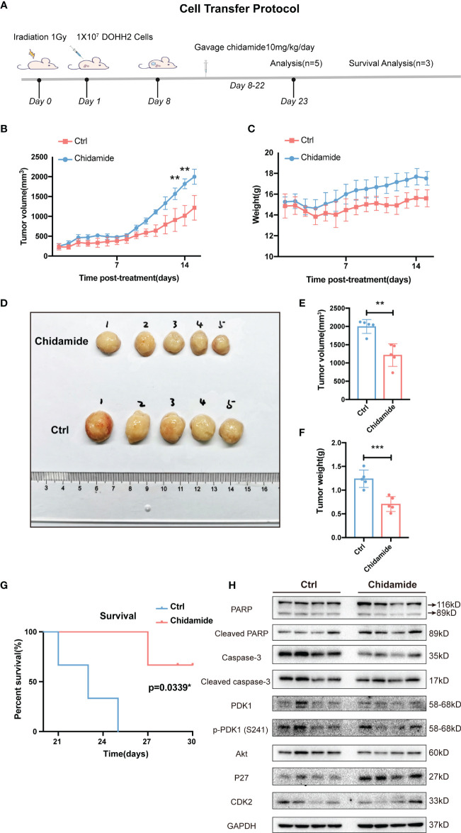
Figure 7.
Effect of chidamide on tumor growth in xenograft mouse models. (A) Cell injection protocol in a FL tumor xenograft model. Tumor volumes (B) and body weights (C) of mice were measured daily and presented as mean ± S.D. (D) Images of tumors from DOHH2-bearing xenograft mice after the indicated treatments (n=10). Tumor volumes (E) and weights (F) in the control and chidamide groups were compared to evaluate the treatment response to chidamide. (G) Kaplan Meier overall survival (OS) curves of tumor-bearing xenograft mice. (H) Chidamide suppressed the PDK1-Akt-P27-CDK2 signaling pathway in vivo. The protein levels of PDK1, AKT, P27, CDK2, PARP, cleaved-PARP, caspase3 and cleaved-caspase-3 were determined by Western blot. (**p < 0.01; ***p < 0.001).

