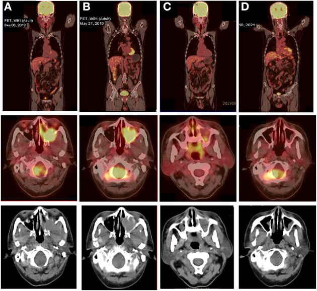
Figure 3.

18F-FDG PET/CT images of the PBL patient at various stages of treatment. (A) Transverse PET/CT scan showed a markedly FDG-avid mass at the left maxillary sinus that invaded the left nasal cavity, left orbital apex, and left alar mandibular space at diagnosis (SUVmax, 7.8; SUVmean, 6.3). (B) After five cycles of V-CHOP, re-evaluating PET/CT showed diminished range of lesions to 3.8 cm × 2.9 cm × 3.8 cm and reduced 18F-FDG uptake in the left maxillary sinus (SUVmax, 7.0; SUVmean, 4.5) and other locations (SUVmax, 4.0; SUVmean, 3.6). (C) After local radiotherapy and two cycles of RV-CDOP, PET/CT showed a complete metabolic response (cMR) with a diminished range of lesions to 1.0 cm × 1.3 cm and significantly reduced 18F-FDG uptake in the left maxillary sinus (SUVmax, 3.5; SUVmean, 2.5) and a lack of metabolic activity in other regions. (D) Follow-up PET-CT 16 months post ASCT showed continued cMR.
