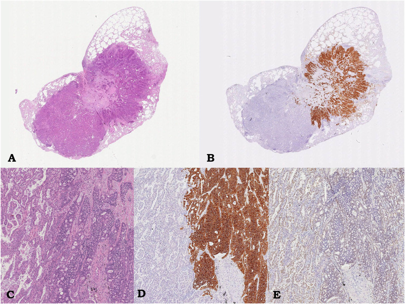
FIGURE 1.
Case 1, pulmonary lesion. Intraparenchymal neoplasm consisting of glandular component corresponding to lepidic-papillary pulmonary adenocarcinoma, on the left, and of adhered trabecular proliferation of monomorphic epithelioid cells corresponding to atypical carcinoid, on the right; hematoxylin/eosin, 0.5x (A) and 5.5x (C). Immunostaining showing diffuse and strong immunoreactivity for Synaptophysin only in the carcinoid component (on the right, 5.5x; B, D) and immunoreactivity for TTF-1 in both component (5.5x, D).

