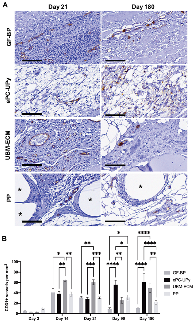
Figure 2. Endothelial cells and neovascularization in GF-BP, ePC-UPy, UBM-ECM, and PP.

(A) GF-BP, ePC-UPy, UBM-ECM, and PP at days 21, and 180 (B) Quantification of CD31+ blood vessels per mm2 of material. With GF-BP, CD31+ endothelial cells were present scattered along the interface of GF-BP implant with surrounding tissue beginning early. However, microvessels with intraluminal RBC were found only on the edges of the material at 180 days post-implantation. At 14 days post-implantation, CD31+ endothelial cells were identified within the ePC-UPy material consistent with neovascularization. By 90 and 180 days, a microvasculature that contained intraluminal RBC was noted within the ePC-UPy, suggesting continuity of these blood vessels with the surrounding host tissue. Abundant neovascularization of the UBM-ECM scaffold was observed early after implantation as indicated by CD31 immunolabeling. Although CD31+ cells were found in the surrounding cellular infiltrate of the PP mesh beginning at 14 days, neovascularization was not present outside of the dense cell layer encapsulating the fibers. All stained for CD31, scale bars 100μm.
