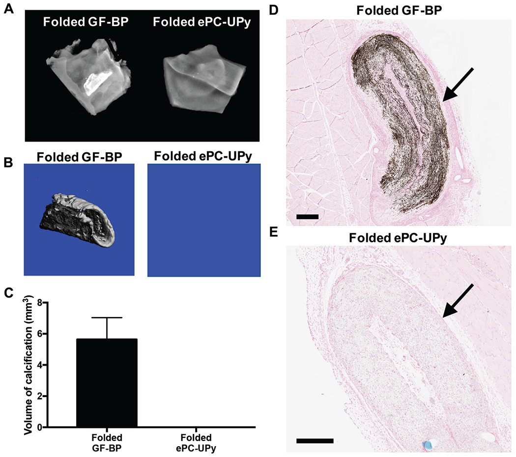
Figure 5. Susceptibility to mechanical stress induced calcification of GF-BP and ePC-UPy, as indicated by intentionally folded specimens.

(A) Representative radiographic images of calcification within folded test materials at 21 days. (B) Representative μCT scans of calcified areas within folded test materials. (C) Quantification of the volume of calcific nodules within each test material as determined by μCT analysis. (D) Mineralization within the folded GF-BP at 21 days as indicated by von Kossa. (E) Lack of mineralization within the folded ePC-UPy. Thus, the high susceptibility to mechanical stress-induced calcification in GF-BP was absent in ePC-UPy. (D) and (E) von Kossa stain, scale bars 400μm.
